
Echocardiogram Test: Types, Procedure, Results & Heart Health Guide
Learn what an echocardiogram is, its types, procedure, and results. Discover how this non-invasive heart ultrasound helps diagnose heart disease and monitor cardiac health.
What is an Echocardiogram? A Complete Guide
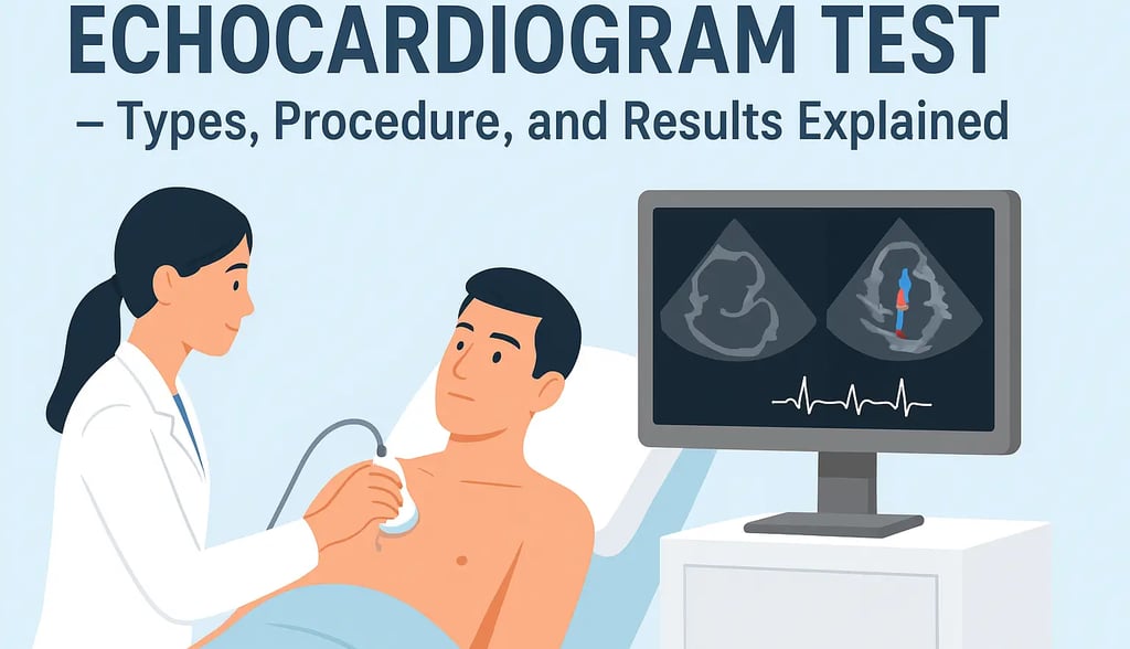
An echocardiogram (often called an echo) is a non-invasive heart imaging test that uses ultrasound technology to create detailed, real-time pictures of the heart. By sending high-frequency sound waves through a handheld device known as a transducer, doctors can see how the heart’s chambers, valves, and blood vessels are functioning.
This test plays a vital role in diagnosing cardiovascular conditions, monitoring heart health, and guiding treatment plans.
How an Echocardiogram Works
During the procedure, a water-based gel is applied to the chest to improve sound wave transmission. The technician then moves the transducer across the chest to capture images from different angles. These images allow cardiologists to:
Evaluate heart chambers
Assess valve function
Monitor blood flow
Detect structural abnormalities
Types of Echocardiograms
Different types of echocardiograms are used depending on the patient’s condition:
Transthoracic Echocardiogram (TTE)
The most common type.
The transducer is placed on the chest wall to capture heart images.
Transesophageal Echocardiogram (TEE)
A special probe is passed down the esophagus.
Provides clearer images when TTE is inconclusive.
Useful for detecting blood clots, infections, or valve problems.
Stress Echocardiogram
Combines ultrasound imaging with exercise or medication.
Helps evaluate heart performance under stress.
Often used to detect coronary artery disease.
Indications for an Echocardiogram
Doctors recommend an echocardiogram for patients showing symptoms of heart problems or when a more detailed evaluation is required. Common reasons include:
Shortness of breath – may indicate heart failure or poor pumping function.
Chest pain – could be linked to coronary artery disease.
Unexplained fatigue – sometimes related to heart dysfunction.
Heart murmurs – to detect valve narrowing or leakage.
Valve diseases – such as aortic stenosis or mitral regurgitation.
Cardiomyopathy – to distinguish between dilated, hypertrophic, or restrictive types.
Congenital heart defects – to identify and monitor structural abnormalities.
Echocardiogram Procedure: What to Expect
An echocardiogram is safe, painless, and requires little preparation. Here’s what typically happens:
Wear comfortable clothing and avoid jewelry on the chest.
You’ll lie on an exam table while a technician applies gel to your chest.
The transducer is moved across your chest to capture images.
The test usually lasts 30–60 minutes.
Some patients may feel slight pressure from the transducer, but discomfort is minimal.
Echocardiogram Results and Interpretation
After the test, a cardiologist reviews the images and measurements. Key findings may include:
Ejection Fraction (EF):
Normal range: 55%–70%.
Lower values may indicate heart failure.
Chamber size and wall motion: Detects enlarged chambers or weak muscle movement.
Valve condition: Checks for narrowing (stenosis) or leakage (regurgitation).
Blood flow patterns: Identifies abnormal circulation or clots.
If abnormalities are found, further tests such as cardiac MRI, CT scans, or blood tests may be recommended.
Next Steps and Treatment
Based on echocardiogram results, treatment may involve:
Lifestyle changes (diet, exercise, quitting smoking)
Medications (to manage blood pressure, heart failure, or arrhythmias)
Surgical procedures (such as valve repair/replacement or bypass surgery)
Regular monitoring with echocardiograms helps track progress and adjust treatment as needed.
Conclusion
An echocardiogram is an essential heart test that provides detailed insights into the heart’s structure and function without surgery or radiation. Whether used for diagnosing conditions, monitoring treatment, or evaluating symptoms, echocardiography plays a crucial role in modern cardiology.
If you experience chest pain, shortness of breath, fatigue, or irregular heartbeat, consult a cardiologist. Early diagnosis with an echocardiogram can make a significant difference in heart health and long-term outcomes.